Volume 38 Number 2 | April 2024
Mahesh Percy, MBA, DMLT; Alvin Billey, MBBS; and Ewarld Marshall, MD, MScMED

Abstract
In hereditary elliptocytosis (HE), the characteristic finding is elliptical red blood cells (RBCs) on the peripheral smear. The patients are usually asymptomatic and rarely present with hemolysis. Because of the silent nature of this condition, HE is difficult and challenging to diagnose. In this article we are presenting an incidental finding of HE during a blood work-up for a patient who was presented to outpatient clinic with fever and fatigue.
Introduction
Hereditary elliptocytosis is a red cell membrane disorder (genetic disorder) that affects the shape of red blood cells. In this condition, the red blood cells have an elliptical or oval shape, instead of their normal round biconcave shape. This abnormal elliptical shape makes the cells more fragile and prone to lysis, which can lead to anemia. The frequency of this disorder is 1 in 5,000 individuals in the United States. This case is an incidental diagnosis during a work-up for fever, mild leukopenia, and a relative lymphocytosis, which supported a diagnosis of a likely viral infection.
Case report
An 11-year-old male presented to the clinic with history of fever and fatigue. The physician at the clinic sends a blood sample for a complete blood count (CBC) work-up and routine chemistries. Laboratory results are shown on this page.
Discussion
Hereditary elliptocytosis is a rare genetic disorder that affects the shape of red blood cells, leading to anemia and other complications. Elliptocytosis was first discovered by Dresbach in 1904 and the hereditary nature was recognized in 1934 by Hunter. This disorder is caused by mutation in genes encoding for proteins involved in maintaining the shape and flexibility of red blood cells. In HE the most common defect is the mutation in alpha or beta chains. Individuals who are heterozygous for an elliptocytic variant are asymptomatic, and individuals with the homozygous variant experience mild to severe anemia. The diagnostic criteria is more than 25 percent of elliptocytes in the peripheral smear.
The clinical presentation of hereditary elliptocytosis can vary from mild to severe anemia, jaundice, and splenomegaly. Some people with hereditary elliptocytosis may have no symptoms and may be diagnosed accidentally during routine blood work.
Diagnosis of hereditary elliptocytosis is usually made by peripheral smear examination which shows the characteristic elliptical or oval shape. Genetic testing can be used to confirm the diagnosis.
Conclusion
Hereditary elliptocytosis is a rare genetic disorder, mostly asymptomatic, that affects the shape of red blood cells. The diagnosis can be confirmed by peripheral smear examination.
Treatment for hereditary elliptocytosis is mainly supportive and involves iron and folic acid supplements to improve anemia. Blood transfusion when severe anemia exists is an alternative option. Genetic counseling may also be recommended for affected individuals and families to help understand the inheritance pattern and mitigate the prevalence of the condition.
References
- Kim, D (24 May 2006). “Elliptocytosis, Hereditary.” Medscape. WebMD LLC. Retrieved 12 August 2013.
- Fathima JL, Sitalakshmi S. Case report on Hereditary Elliptocytosis. Ind J Med Case Rep 2013; 2(2): 41-43.
- Greenberg LH, Tanaka KR. Hereditary elliptocytosis with hemolytic anemia-a family study of five affected members. Calif Med 1969; 110(5): 389-93.
- Weiss HJ: Hereditary elliptocytosis with haemolytic anemia, Am J Med 1963; 35:455-466.
- PS Sharmila, Kannupriya, MF Paul. Hereditary Elliptocytosis. The journal of Medical Sciences. 2015; 1. 41-3.
- Hoffman, R; Benz, E; Shattil, S; Furie, B; Cohen, H (2005). Hoffman Hematology: Basic Principles and Practice (4th ed.). Philadelphia: Churchill Livingstone. ISBN 978-0-443-06628-3.
- Bannerman, Rm; Renwick, Jh (July 1962). “The hereditary elliptocytoses: clinical and linkage data.” Annals of Human Genetics. 26 (1): 23–38. doi:10.1111/j.1469-1809.1962.tb01306.x. ISSN 0003-4800. PMID 13864689. S2CID 40636103
- Tse, Wt; Lux, Se (January 1999). “Red blood cell membrane disorders.” British Journal of Haematology. 104 (1): 2–13. doi:10.1111/j.1365-2141.1999.01130.x. ISSN 0007-1048. PMID 10027705.
- Warang P, Kedar P. Hereditary Elliptocytosis: A Rare Red Cell Membrane Disorder. Indian J Hematol Blood Transfus. 2018 Oct;34(4):754-755. [PMC free article] [PubMed]
- Dresbach M (1904). “Elliptical human red corpuscles.” Science. 19 (481): 469–470. Bibcode:1904Sci….19..469D. doi:10.1126/science.19.481.469. PMID 17730874.
- Gallager PG. Hereditary Elliptocytosis: Spectrin and protein 4.1R. Semin hematol. 2004; 41; 142-64.
- Hoffbrand AV, Catovsky D, Tuddenham EGD and Green AR (2011). Hereditary disorders of red cell membrane In Postgraduate Haematology 6. Wiley-Blackwell Publications.
- Bannerman, R M; Renwick, J H. The hereditary elliptocytoses: clinical and linkage data. Annals of human genetics 26: 23-38.
Mahesh Percy is an Instructor/Medical Technologist at St. George’s University in St. George’s, Grenada.
Alvin Billey is a Lecturer, Associate College Director at St. George’s University in St. George’s, Grenada.
Ewarld Marshall is Chair, Pathology at the Medical Pathology Diagnostic Lab Director at St. George’s University in St. George’s, Grenada.
This article was previously published by the Caribbean Association of Medical Technologists in their October 2023 BGM and Scientific magazine.
Hematological tests
Hemoglobin
RBC
Hematocrit
MCV
MCH
MCHC
RDW
MPV
WBC
Neutrophils
Lymphocytes
Eosinophils
Monocytes
Basophils
Platelets
Patient value
12.7 g/dl
4.37 10*6/ul
36.8%
84.3 fl
29.0 pg
34.4 g/dl
12.2%
7.5 fl
3.5 10*3/ul
27.4%
63.5%
2.6%
6.1%
0.4%
371 10*3/ul
Normal value
14 – 18 g/dl
4.5 – 6.2 10*6/ul
40 – 54%
82 – 98 fl
26 – 34 pg
31 – 38 g/dl
11.6 – 13.7%
7.8 – 11.0 fl
4.5 – 11.0 10*3/ul
40 – 70%
20 – 40%
1 – 4%
2 – 8%
0 – 1%
150 – 400 10*3/ul
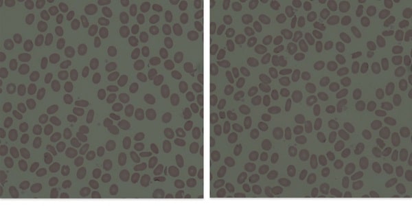
The peripheral smear examination showed elliptical or oval shaped red blood cells (more than 25 percent) with occasional polychromatic cells, spherocytes, and schistocytes. The findings suggest hereditary elliptocytosis.
Biochemistry tests
Fasting Blood Sugar
BUN
Creatinine
Total bilirubin
Direct bilirubin
SGOT/AST
SGPT/ALT
Alkaline Phosphatase
Patient value
70.7 mg/dl
10.7 mg/dl
0.8 mg/dl
0.50 mg/dl
0.17 mg/dl
24.7 U/L
22.9 U/L
376.7 U/L (In children the Alkaline Phosphatase levels may be elevated until epiphysis close)
Normal value
60 – 110 mg/dl
7 – 18 mg/dl
0.4 – 1.4 mg/dl
0.2 – 1.2 mg/dl
0.0 – 0.30 mg/dl
5 – 34 U/L
4 – 36 U/L
35 – 123 U/L
Total protein
Albumin
Total cholesterol
HDL
LDL
VLDL
Triglycerides
Sodium
Potassium
Chloride
7.65 g/dl
4.6 g/dl
153 mg/dl
46.6 mg/dl
89.0 mg/dl
17 mg/dl
97.0 mg/dl
139.4 mmol/L
3.91mmol/L
105.1 mmol/L
6.6 – 8.8 g/dl
3.5 – 5.2 g/dl
135 – 200mg/dl
>40 mg/dl
<130 mg/dl
0 – 40 mg/dl
60 – 165 mg/dl
135 – 155 mmol/L
3.6 – 5.5 mmol/L
96 – 106 mmol/L